Aesthetic Medicine & 3D Imaging Blog
Latest clinical research, technical advances, and practice growth insights from LifeViz®.
5 Best Breast Enhancement 3D Imaging Systems?
Table of Contents
- Clinical Demand: ASPS Recorded 304,000 U.S. Breast Augmentations in 2023
- 3D Imaging Fundamentals: Structured Light, Photogrammetry, and Millimeter Accuracy
- Five Leading Platforms: VECTRA, Crisalix, Axis Three, 3dMD, and Arbrea
- Evidence-Based Comparison: Capture Time, Repeatability, and Simulation
- Safety and Selection: FDA 510(k), CE MDR, HIPAA, and Data Security
- FAQS
- Conclusion
- Related Posts
Choosing the 5 Best Breast Enhancement 3D Imaging Systems requires more than comparing attractive screen images. These platforms can capture a patient’s torso, map breast volume, and simulate possible enhancement outcomes. A clear scan may show asymmetry that is difficult to notice in a mirror. That matters.
This guide examines systems through clinical usefulness, image quality, workflow, and patient communication. It considers how quickly each platform captures images, how naturally it displays body contours, and whether surgeons can explain limitations without creating unrealistic expectations. A reliable system should support consultation, not replace physical examination, medical history, or professional judgment. Results vary.
Experience also matters. A skilled clinician may interpret a modest difference in projected volume more responsibly than an impressive digital preview. The best Breast Enhancement 3D Imaging technology should help patients ask informed questions about proportions, implant options, tissue characteristics, and possible risks. It should also protect sensitive photographs through secure storage and controlled access.
No system is perfect. Simulations can differ from surgical outcomes because healing, swelling, skin elasticity, implant position, and individual anatomy change the final appearance. This article therefore avoids treating software rankings as universal truth. Instead, it compares practical strengths and weaknesses, including learning curves, image consistency, consultation value, and the quality of supporting evidence. Readers should verify regulatory status, clinical credentials, and privacy policies before selecting a provider. A thoughtful decision may feel less exciting than a dramatic preview, but it is usually more trustworthy.
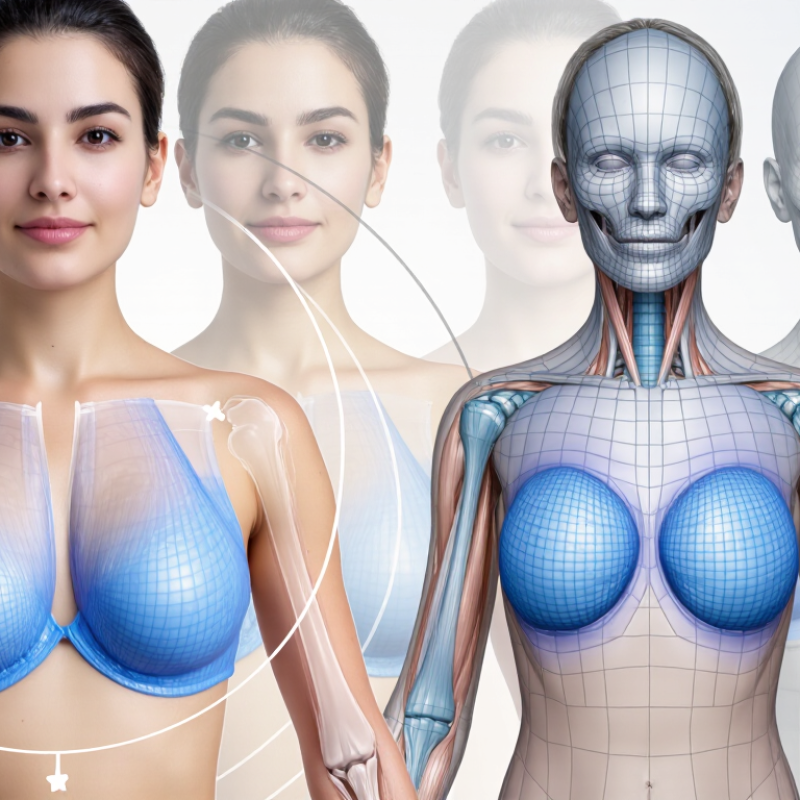
Clinical Demand: ASPS Recorded 304,000 U.S. Breast Augmentations in 2023
The demand for breast augmentation keeps clinical visualization relevant. The American Society of Plastic Surgeons recorded 304,000 breast augmentations in the United States in 2023. That volume gives consultation technology a serious role, not a decorative one.
The five strongest 3D imaging systems should offer accurate body capture, fast processing, clear implant simulations, secure data handling, and simple clinical documentation. A patient may stand before a camera for less than a minute. The screen can then show front, oblique, and side views. That visual conversation often reveals differences between desired shape, implant volume, and realistic tissue limits. However, simulation is not a promise. The American Society of Plastic Surgeons emphasizes that outcomes depend on anatomy, technique, healing, and implant selection.
Professional evaluation should also consider evidence quality. The International Society of Aesthetic Plastic Surgery continues to report breast augmentation as a major global aesthetic procedure. Meanwhile, the U.S. Food and Drug Administration requires detailed implant information and patient risk communication. A useful imaging platform should support those discussions, rather than replace them.
Some systems may appear impressive but produce weak results under uneven lighting or unusual anatomy. That matters. Clinicians should compare simulated images with postoperative photography, patient feedback, and documented follow-up. The best tool is not always the most visually dramatic. It is the one that improves understanding without creating false certainty.
U.S. Breast Procedure Demand in 2023
Breast augmentation remained a major elective procedure category, with approximately 304,000 procedures recorded in the United States in 2023.
The chart presents rounded 2023 U.S. procedure volumes reported by the American Society of Plastic Surgeons. These demand levels help explain why 3D imaging is increasingly used to support patient education, surgical planning, and expectation management for breast enhancement procedures.
3D Imaging Fundamentals: Structured Light, Photogrammetry, and Millimeter Accuracy
5 Best Breast Enhancement 3D Imaging Systems?
Structured light projects a patterned grid across the torso. Cameras then measure surface changes from grid distortion. It is fast, repeatable, and useful in controlled rooms. Photogrammetry builds a three-dimensional model from many overlapping photographs. It can capture natural posture more comfortably, but lighting and movement can reduce reliability. Hybrid systems combine both approaches. The best choice depends on workflow, patient comfort, and measurement goals.
Millimeter accuracy sounds impressive, but accuracy needs context. A 1 mm difference may reflect posture, breathing, or camera placement. Published validation studies in aesthetic imaging commonly report repeatability within a few millimeters, rather than perfect anatomical truth. The International Society of Aesthetic Plastic Surgery reported more than one million breast augmentation procedures worldwide in its 2023 survey. That volume increases the value of consistent documentation, but imaging cannot replace physical examination. The American Society of Plastic Surgeons also reported 304,181 breast augmentation procedures in the United States in 2023. These figures show demand, not proof of surgical outcomes.
Tips: Use the same room, lighting, stance, and arm position every time. Mark anatomical landmarks carefully. Record the device’s calibration status. Compare scans with clinical findings, not alone. A useful image should be repeatable, understandable, and clinically relevant. This is where practice can expose weaknesses. Even skilled operators may create small measurement errors. Teams should review those errors openly and avoid promising millimeter-level certainty without validation data.
Five Leading Platforms: VECTRA, Crisalix, Axis Three, 3dMD, and Arbrea
Breast enhancement imaging has moved beyond glossy before-and-after galleries. The five leading platforms use three-dimensional capture, anatomical landmarks, and virtual sizing. Their value is practical: patients can inspect projected width, profile, and asymmetry before surgery. In clinic, consistent lighting and posture matter as much as software. A tilted shoulder can distort the result.
The systems differ in workflow. One emphasizes high-resolution photographic capture. Another offers browser-based consultations and rapid implant simulations. A third focuses on surface mapping, while others support volumetric analysis or augmented-reality viewing. Surgeons should compare calibration, export options, data protection, and training requirements. A beautiful rendering is not clinical evidence.
The American Society of Plastic Surgeons reported more than 300,000 breast augmentation procedures in the United States during 2023. The International Society of Aesthetic Plastic Surgery also listed breast augmentation among the world’s leading surgical aesthetic procedures in its 2023 global survey. These figures increase the need for clearer consent conversations, not stronger sales language.
Accuracy remains imperfect. Skin elasticity, healing, implant movement, and surgical technique can change the final appearance. A 3D preview is a planning aid, not a promise. Independent validation should include varied skin tones, body shapes, and repeat scans. That part is often underexplained. Professional judgment still matters.
Evidence-Based Comparison: Capture Time, Repeatability, and Simulation
5 Best Breast Enhancement 3D Imaging Systems?
A practical comparison should begin with capture time, not glossy simulation images. Multi-camera systems often capture a full breast surface in under one second. Structured-light systems commonly require two to five seconds. Handheld depth scanners may take longer, especially around the inframammary fold. Motion matters. A patient’s breathing can create visible contour shifts.
Published validation studies in Aesthetic Surgery Journal and Plastic and Reconstructive Surgery frequently report repeatability above 0.95 using intraclass correlation coefficients. Some studies also report surface measurement errors below 1 millimeter under controlled conditions. These results are encouraging, but clinics rarely operate under laboratory conditions. Hair, skin reflectivity, posture, and inconsistent lighting can reduce reliability.
The five most useful system categories include
multi-camera stereophotogrammetry, structured-light scanning, handheld depth capture, smartphone-assisted imaging, and hybrid systems. Multi-camera platforms usually offer the fastest workflow. Structured light can provide detailed surface geometry. Handheld devices may fit smaller practices, but operator technique becomes more influential. Smartphone systems are convenient, yet independent validation remains thinner.
Simulation quality deserves caution. A 2023 professional consensus report on digital planning emphasized that visual prediction depends on tissue behavior, implant position, healing, and patient anatomy. In other words, simulation is a communication tool, not a promise. My own concern is simple: a beautiful render can hide weak repeatability. Clinics should request test-retest data, raw measurement accuracy, and documented capture conditions before trusting any “best” list.
Safety and Selection: FDA 510(k), CE MDR, HIPAA, and Data Security
5 Best Breast Enhancement 3D Imaging Systems?
Safety and Selection: FDA 510(k), CE MDR, HIPAA, and Data Security
Choosing among five leading 3D breast imaging systems requires more than comparing image resolution. Start with intended use, clinical evidence, operator training, and patient comfort. A useful system should produce consistent images under real clinic conditions, including uneven lighting, motion, and different body types. Small details matter. Ask to review validation data, known limitations, and examples of image artifacts.
FDA 510(k) clearance indicates substantial equivalence to a legally marketed device; it is not a universal safety guarantee. In Europe, CE MDR conformity reflects documented safety, performance, and post-market obligations. These pathways differ. A serious evaluation should verify the exact model, intended indications, software version, and responsible legal manufacturer. Local regulatory requirements may also apply. Do not rely on a sales presentation alone.
Privacy deserves equal attention. If a system handles identifiable patient images in the United States, determine whether HIPAA applies and whether a business associate agreement is required. Confirm encryption during transfer and storage, role-based access, multifactor authentication, audit logs, retention controls, and secure deletion. Ask where cloud data is stored and how incidents are reported. Qualified clinicians, biomedical engineers, and privacy officers should test the workflow together. A polished demonstration can hide practical weaknesses. Even a careful checklist can miss them. Reassess security after updates, staff changes, and integration with electronic health records.
5 Best Breast Enhancement 3D Imaging Systems? – Safety and Selection: FDA 510(k), CE MDR, HIPAA, and Data Security
Comparison of five clinically relevant 3D imaging approaches for breast enhancement consultation, planning, documentation, and follow-up. Regulatory and security status must be verified for the exact device, software version, intended use, and healthcare deployment.
| Imaging System Type | Best Clinical Use | Patient Exposure and Safety | Typical 3D Output | FDA 510(k) Selection Check | CE MDR Selection Check | HIPAA and Privacy Considerations | Minimum Data-Security Controls |
|---|---|---|---|---|---|---|---|
|
1. Optical 3D Surface Imaging
Radiation-free
|
Baseline consultation, implant or augmentation simulation, body-surface documentation, and postoperative visual comparison. | Uses visible or near-infrared light and does not use ionizing radiation. It does not replace diagnostic imaging for suspicious masses or tissue pathology. | Textured or non-textured surface mesh, digital measurements, symmetry analysis, and simulated contour visualization. Usually limited to externally visible anatomy. | Confirm whether the exact system is marketed as a medical device in the United States and whether its intended use requires FDA clearance. Check the FDA 510(k) database and labeling; cosmetic visualization claims may differ from diagnostic claims. | Confirm a valid CE marking under Regulation (EU) 2017/745 where applicable, the device classification, Declaration of Conformity, and current manufacturer documentation. | Facial and breast surface images can be identifiable health information when linked to a patient record. Use a HIPAA-compliant workflow when operated by a covered entity or business associate. | Encryption in transit and at rest, role-based access, multifactor authentication, audit logs, controlled exports, retention limits, secure deletion, and documented breach-response procedures. |
|
2. Multi-View Stereophotogrammetry
Radiation-free
|
High-detail preoperative planning, asymmetry assessment, patient education, and longitudinal photographic documentation. | Uses synchronized cameras and controlled lighting without ionizing radiation. Accuracy depends on patient posture, breathing, motion, lighting, and camera calibration. | Full-color 3D surface model, point cloud or polygon mesh, linear and volumetric measurements, and side-by-side simulation views. | Verify the precise medical intended use, predicate comparison, software functions, and cleared indications in the official 510(k) record. Do not assume that a camera-based system is cleared for diagnostic interpretation. | Verify the applicable MDR classification, conformity-assessment route, notified-body information where required, post-market surveillance documentation, and instructions for use. | Store images and derived meshes as protected health information when linked with identifiers. Obtain appropriate consent for simulation images, teaching use, research, and marketing. | Use encrypted acquisition and transfer, unique user accounts, least-privilege permissions, MFA, immutable audit trails, secure cloud configuration, backup testing, and export watermarking where appropriate. |
|
3. Structured-Light 3D Scanning
Radiation-free
|
Fast surface capture for standardized measurements, posture-controlled comparisons, and integration with digital planning software. | Projects patterned light onto the skin and generally uses non-ionizing optical energy. Eye-safety documentation and operating-distance instructions should be reviewed. | High-density surface mesh, texture map, landmarks, distance measurements, symmetry reports, and exportable 3D files such as OBJ, PLY, or STL. | Determine whether the device and connected planning software are regulated separately or as a system. Confirm FDA clearance status, intended-use wording, software version, and any limitations on patient-specific recommendations. | Confirm CE MDR status for the complete configuration, including scanner, software, accessories, and cloud services if supplied as part of the medical-device system. | 3D meshes may remain identifiable even after obvious facial features are removed. Apply data minimization, patient authorization rules, access controls, and documented retention schedules. | Require TLS-protected connections, strong encryption at rest, secure API authentication, vulnerability-management processes, software-update controls, audit logging, and tested disaster recovery. |
|
4. 3D Ultrasound
Diagnostic adjunct
|
Assessment of superficial tissue, implant-related findings, fluid collections, and selected follow-up questions when clinically indicated. | Uses non-ionizing sound waves. It is operator-dependent and should follow the ALARA principle: use the lowest acoustic output and shortest exposure that reasonably achieves the clinical objective. | Volumetric ultrasound data, multiplanar reconstructions, cine loops, caliper measurements, and structured imaging reports. Image quality varies with tissue depth and operator skill. | Verify an FDA-cleared ultrasound platform and compatible transducer for the intended application. Review thermal and mechanical index displays, acoustic-output information, software indications, and service requirements. | Confirm CE MDR documentation for the ultrasound console, probes, software, and accessories. Check cleaning, disinfection, and reprocessing instructions for reusable transducers. | DICOM studies, reports, and patient identifiers are protected health information in an applicable clinical workflow. Use authenticated PACS connections and formal user-access management. | DICOM over secure network transport where supported, encryption at rest, role-based PACS access, MFA, audit trails, endpoint protection, patch management, and validated backup and restore procedures. |
|
5. MRI-Based 3D Breast Imaging
Advanced diagnostic imaging
|
High-detail diagnostic evaluation, implant integrity assessment in appropriate protocols, complex asymmetry, and cases requiring soft-tissue characterization. | Uses a strong static magnetic field and radiofrequency energy rather than ionizing radiation. Strict screening is required for implants, foreign bodies, devices, pregnancy considerations, claustrophobia, and contrast-related risks. | Three-dimensional cross-sectional image volumes, multiplanar reconstructions, maximum-intensity projections, quantitative measurements, and DICOM-compatible studies. | Confirm FDA clearance for the complete MRI system and applicable coils, sequences, software, and contrast-related accessories. Review MR safety labeling, field strength, implant conditions, and intended-use statements. | Verify CE MDR conformity for the scanner configuration and associated software, plus documented MR safety information, clinical evaluation, post-market surveillance, and local installation requirements. | MRI examinations contain direct identifiers and sensitive clinical findings. Use controlled PACS access, minimum-necessary disclosure, patient identity verification, and documented authorization for secondary use. | Secure DICOM transfer, encrypted storage, network segmentation, MFA, role-based access, comprehensive audit logs, vendor remote-access controls, tested backups, and formal incident-response procedures. |
FAQS
It should capture body shape quickly, create repeatable images, and support clear patient discussions. A patient may stand before several cameras for less than one minute. The system should show front, angled, and side views. Visual clarity matters, but it cannot replace clinical judgment.
Common options include multi-camera capture, structured light, handheld depth scanning, smartphone-assisted imaging, and hybrid systems. Multi-camera systems usually provide fast workflows. Structured light can show detailed surface geometry. Handheld systems may suit smaller clinics, but operator technique matters more.
Multi-camera systems may capture a full surface in under one second. Structured-light systems often need two to five seconds. Handheld devices can take longer around the lower breast fold. Breathing matters.
Controlled studies often report repeatability above 0.95. Some report surface errors below one millimeter. Real clinics are less predictable. Hair, skin reflection, posture, breathing, and uneven lighting can reduce reliability. The numbers may look better than everyday performance.
No simulation can guarantee the final appearance. Results depend on anatomy, tissue behavior, implant position, surgical technique, healing, and selected volume. A simulation is a communication tool. It helps compare expectations with realistic tissue limits. A beautiful render can still create false confidence.
Ask for test-retest data, measurement accuracy, capture conditions, and known limitations. Request postoperative comparisons and documented patient feedback. Check performance with different body types and lighting conditions. Do not judge quality from glossy images alone. That shortcut is tempting.
Verify the exact model, intended use, software version, and responsible legal manufacturer. Check applicable clearance, conformity, and local regulatory requirements. Confirm operator training and clinical documentation procedures. A sales demonstration may not reveal workflow problems. Testing matters more.
Use encryption during transfer and storage, role-based access, multifactor authentication, and audit logs. Confirm retention periods, secure deletion, cloud storage locations, and incident reporting procedures. Determine whether healthcare privacy rules require additional agreements. Review security after software updates and staff changes. Small gaps remain possible.
Conclusion
Breast Enhancement 3D Imaging is becoming an important tool in modern consultation and treatment planning, driven by the high demand for breast augmentation procedures in the United States. These systems use technologies such as structured light and photogrammetry to capture detailed three-dimensional body images, often with millimeter-level accuracy. By creating realistic visual simulations, they help patients and clinicians discuss expectations, compare possible outcomes, and support more individualized planning.
This overview examines five leading categories of clinical platforms without focusing on specific brands, comparing them by capture speed, repeatability, image quality, and simulation capabilities. It also highlights essential selection criteria, including regulatory clearance or certification, patient privacy, HIPAA-aligned practices, data encryption, and secure storage. While 3D imaging cannot guarantee surgical results, evidence-based evaluation and careful clinical interpretation can improve communication, decision-making, and patient confidence. The most suitable system should balance technical performance, workflow efficiency, usability, and strong protection of sensitive medical information.

Blog Tags: