Knowledge & Innovation

Aesthetic Medicine & 3D Imaging Blog

Latest clinical research, technical advances, and practice growth insights from LifeViz®.

0%

In 2026, body contouring decisions increasingly depend on measurable, three-dimensional information. A Body Contouring 3D Assessment can show body shape, volume changes, and surface symmetry more clearly than standard photographs. However, impressive images do not automatically mean reliable results. The technology must support qualified professionals, informed patients, and realistic treatment planning.

Choosing the right system requires more than comparing camera resolution. Consider scanning speed, measurement consistency, software usability, and compatibility with clinical workflows. Ask whether the device has documented validation, regular calibration procedures, and transparent limitations. A useful system should produce repeatable measurements in ordinary conditions, even when lighting, posture, or skin texture changes slightly. The details matter.

Privacy also deserves serious attention. Patient images and body measurements should be collected with informed consent, protected through appropriate security controls, and handled according to applicable privacy requirements. Clinics should examine data storage, access permissions, export options, and vendor support before purchase. These practical questions often reveal more than a polished product demonstration.

Experience remains important. A skilled practitioner can interpret three-dimensional data while recognising swelling, posture effects, and measurement errors. No system replaces clinical judgement. Accuracy is not everything. Cost, training time, patient communication, and long-term maintenance also influence value. Some platforms may appear advanced but offer limited evidence or complicated reports. That weakness should be acknowledged, not hidden. This guide explains how to compare Body Contouring 3D Assessment solutions in 2026, using practical performance, professional expertise, ethical safeguards, and credible evidence as the main decision standards.
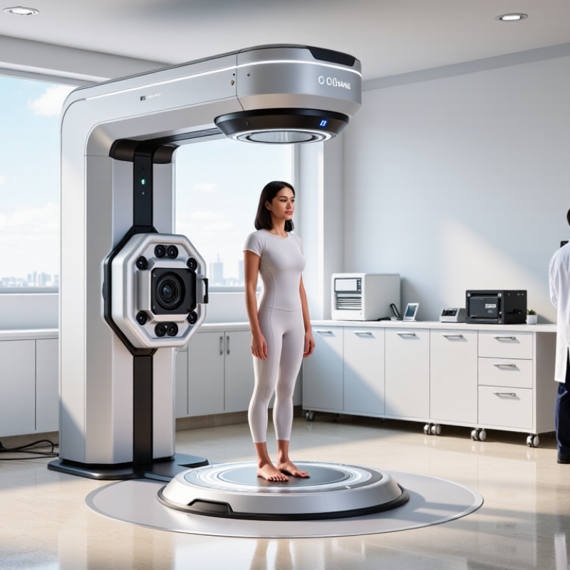
How to Choose Body Contouring 3D Assessment in 2026?

What Is 3D Body Contouring Assessment?

What Is 3D Body Contouring Assessment?

A 3D body contouring assessment creates a digital model of the body using calibrated cameras or scanners. It captures shape, volume, posture, and surface proportions from several angles. It is not a simple photograph.

During a typical session, the person stands still in consistent lighting and neutral clothing. The system records areas such as the waist, hips, abdomen, thighs, and shoulders. Trained professionals can compare measurements over time and identify changes that ordinary scales may miss. A two-centimeter waist change may matter, even when body weight stays similar. Small differences matter.

Reliable assessment requires careful preparation. The operator should check scanner calibration, body position, lighting, and measurement settings. Privacy and informed consent also deserve attention. A digital model is useful, but it is not a diagnosis or a promise of treatment results. Health history and a physical evaluation remain important.

The process is not flawless. Loose clothing, uneven posture, breathing, or reflective surfaces can affect the scan. A precise-looking image may still contain errors. For this reason, professionals should repeat scans under similar conditions and explain the limits clearly. I would question any report that presents one scan as a complete picture of health. Three-dimensional data can guide realistic goals, but human judgment must remain part of the assessment.

Which Body Measurements Should the System Capture?

A reliable 3D body contouring assessment should capture more than attractive front and side images. It should record height, weight, and body position under consistent conditions. Circumferences matter most when tracked at clear anatomical landmarks. These include the neck, chest or bust, waist, abdomen, hips, upper arms, thighs, and calves. The system should also measure vertical lengths, such as torso height and leg length. Small changes can reveal uneven treatment responses.

Shape data adds important context. Look for shoulder, hip, and waist proportions. Capture left-right differences, posture, spinal alignment, and visible surface contours. A useful system should show depth changes, not only circumference changes. For example, a waist may look smaller from the front but remain unchanged in side depth. That difference can affect clinical decisions and client expectations.

Measurement quality depends on repeatability. The person should stand naturally, with feet positioned consistently and arms slightly away from the body. Lighting, clothing, camera distance, and breathing can alter results. They should be controlled. Ask whether the system provides calibration checks, measurement landmarks, and exportable records. Independent validation is valuable, but it should not replace professional judgment. No scan is perfect. Body movement still creates noise. I would compare repeated scans before interpreting a small change. A two-millimeter difference may reflect posture, not treatment progress. Clear consent, secure data handling, and trained operators also support trustworthy assessment.

How to Compare 3D Assessment Technologies in 2026

How to Choose Body Contouring 3D Assessment in 2026?

Comparing 3D assessment technologies requires more than viewing attractive body images. Start with measurement accuracy, repeatability, and clinical workflow. In practical evaluations, I compare the same person twice, under identical lighting and posture. Small changes in shoulder position can distort circumference readings. That detail is easy to miss.

Camera-based photogrammetry usually offers fast capture and accessible operation. Depth-sensing systems can record body shape with less dependence on image texture. Structured-light systems may produce sharper surface detail, but they often require controlled positioning. Artificial intelligence can improve landmark detection, although its results still depend on training data and technician oversight. No system is perfect. I would question any platform promising identical outcomes for every body type.

Tips: Ask how the system is calibrated, how often accuracy is checked, and whether reports separate measured data from visual estimates. Review sample reports before purchasing. Test people with different heights, skin tones, body shapes, and mobility levels. Check whether the technology tracks changes consistently over time, not just whether one scan looks impressive. Also confirm data protection procedures, staff training, and export options. A reliable assessment should support informed decisions, not replace professional judgment. I still find that rushed operators create avoidable errors. Technology helps, but disciplined practice matters more.

What Factors Affect Accuracy, Privacy, and Usability?

Choosing a body contouring 3D assessment requires more than comparing image quality. Accuracy depends on camera calibration, lighting, distance, body position, and software validation. A bright window can create shadows around the waist. Loose clothing can distort measurements. Consistent scanning conditions matter.

In practice, repeatability is often more useful than a single impressive scan. Ask whether trained operators obtain similar results on different days. Review measurement error, reference populations, and independent validation data. The system should show confidence ranges, not only attractive body maps. No scan is perfectly objective. Human posture still affects the result.

Privacy deserves equal attention. Confirm informed consent, encryption, access controls, and clear data-retention periods. Sensitive body images should not remain indefinitely by default. Check whether users can request deletion or correct inaccurate records. Usability also affects reliability. A confusing interface encourages rushed positioning and missed instructions. Look for guided capture, accessibility features, fast processing, and straightforward export options. Staff should understand the system’s limitations before discussing results with clients. That part is easy to overlook. A short pilot with real users may reveal more than a polished demonstration.

How to Select the Right 3D Assessment for Your Goals

Choosing a 3D body assessment should begin with your goal, not the device. Are you tracking fat reduction, skin laxity, muscle definition, or surgical planning? Each purpose needs different measurements. A full-body scan may show posture and volume changes, while localized imaging can reveal waist asymmetry more clearly. This matters as global demand grows. The ISAPS Global Survey 2023 reported nearly two million liposuction procedures worldwide.

Ask how the system captures evidence. Consistent lighting, posture, distance, and clothing are essential. Otherwise, a three-centimeter waist change may reflect positioning, not treatment. The American Society of Plastic Surgeons’ 2023 Procedural Statistics also listed body contouring among commonly requested aesthetic procedures. That trend increases the need for repeatable assessments. Look for documented measurement accuracy, secure data handling, and trained operators.

Your timeline matters too. Monthly tracking needs fast, comfortable scans. Preoperative planning requires detailed surface mapping and careful clinician review. Do not select software only because its images look impressive. Ask whether it measures your actual target. Skin texture can look better before volume changes appear. Or the reverse. I have seen patients focus on attractive color maps while missing posture differences. That is a useful warning. A reliable assessment should support clinical judgment, not replace an in-person examination. Cite reports directly, record the same poses, and question results that seem too perfect.

How to Choose Body Contouring 3D Assessment in 2026? – How to Select the Right 3D Assessment for Your Goals
Assessment Method Typical Capture Time Typical Measurement Capability Quantitative Outputs Best Suited Goals Main Strengths Important Limitations Relative Cost and Practicality
Multi-view 3D Photogrammetry Usually 1–10 seconds for image capture, followed by automated processing. Commonly approximately 1–3 mm under controlled lighting, positioning, and calibration conditions. Body circumference, surface area, volume estimates, posture symmetry, regional shape changes, and side-by-side surface comparisons. Documenting aesthetic treatment progress, comparing pre- and post-treatment shape, and creating patient-friendly visual reports. Non-contact, fast, visually intuitive, and suitable for repeated follow-up assessments. Results can be affected by loose clothing, hair, reflective surfaces, lighting, pose changes, and incomplete body coverage. Moderate; highly practical
Structured-Light 3D Scanning Approximately 1–5 seconds for a body region or full-body scan, depending on the scanning setup. Often approximately 0.5–2 mm in controlled clinical environments; actual performance depends on calibration and scanning distance. Surface contours, circumference, localized depth, volume, area, symmetry, and three-dimensional deviation maps. High-detail contour analysis, precise regional measurement, and standardized clinical outcome tracking. High geometric detail and strong repeatability when the operator, posture, lighting, and workflow are standardized. Requires controlled capture conditions; movement, occlusion, dark clothing, and reflective materials may reduce data quality. Moderate to high; clinic-based
Depth-Camera or Mobile 3D Capture Usually less than 1–5 seconds for image acquisition, with processing completed in seconds to minutes. Often approximately 3–10 mm for practical body measurements, with accuracy varying by sensor, distance, software, and environment. Approximate circumference, body width, regional volume trends, posture, and visual shape comparisons. Accessible progress monitoring, preliminary consultations, remote workflows, and frequent measurements. Portable, relatively low-cost, quick to deploy, and convenient for repeated assessments. Generally less precise than calibrated clinical scanners; small changes may be hidden by sensor noise or inconsistent positioning. Low to moderate; highly accessible
Handheld 3D Surface Scanner Approximately 1–10 minutes, depending on the body area, operator technique, and required scan density. Commonly approximately 0.5–2 mm for suitable surfaces when the device is properly calibrated and operated. Detailed surface topology, localized contour depth, surface area, volume, and three-dimensional change maps. Small or complex body regions, scar and contour documentation, and targeted pre- and post-treatment analysis. Flexible access to difficult areas and potentially high local surface detail. Operator-dependent; tracking can be lost during movement, and full-body scanning may require multiple scans and data alignment. Moderate to high; skill-dependent
2D Standardized Photography Usually less than 1 minute when camera position, lighting, background, and pose are standardized. No direct depth measurement; apparent size and shape depend strongly on camera angle, distance, lens, and posture. Visual documentation, visible asymmetry, skin appearance, posture, and qualitative shape comparison. Basic documentation, patient communication, and situations where quantitative 3D measurement is not required. Simple, inexpensive, familiar, and easy to archive. Cannot reliably measure true volume or three-dimensional change; perspective and pose can create misleading differences. Low; highly practical
Ultrasound-Based Soft-Tissue Assessment Approximately 5–30 minutes for selected regions, depending on the number of sites and examination protocol. Depth and tissue-thickness measurements can be in the millimeter range, but results depend on the operator, probe pressure, and tissue characteristics. Subcutaneous tissue thickness, skin and soft-tissue layers, localized tissue composition indicators, and targeted anatomical measurements. Evaluating localized soft-tissue thickness when surface shape alone does not explain the treatment outcome. Provides information beneath the skin and avoids ionizing radiation. Not a full-body surface assessment; probe pressure, anatomical site, operator skill, and measurement protocol affect repeatability. Moderate; targeted use
MRI-Based Body Composition Assessment Typically 20–45 minutes, depending on the body region, sequence, and imaging protocol. High anatomical detail with spatial resolution commonly measured in millimeters; exact performance depends on the protocol and scanner. Subcutaneous and visceral fat distribution, muscle volume, tissue cross-sectional area, and internal anatomical structure. Research-grade body-composition analysis, complex anatomical evaluation, and cases requiring internal tissue information. Provides detailed internal anatomy without ionizing radiation and can distinguish multiple soft-tissue compartments. Expensive, less convenient, time-consuming, and generally unnecessary for routine cosmetic surface-progress tracking. High; specialized use
Typical values are indicative ranges reported for commonly used assessment workflows. Actual accuracy and repeatability depend on the device, software, calibration, lighting, operator training, subject movement, clothing, posture, and measurement protocol. For reliable progress tracking, use the same method, settings, body position, and measurement landmarks at every session.

FAQS

What should be compared when evaluating 3D body assessment technologies?

Compare measurement accuracy, repeatability, workflow, data security, and report clarity. Attractive images alone prove very little.

How can repeatability be tested?

Scan the same person twice with identical lighting, posture, clothing, and camera distance. Small shoulder movements can change circumference readings.

What are the differences between common scanning methods?

Camera-based systems capture quickly and operate easily. Depth sensors record shape with less reliance on image texture. Structured light can show sharper surface details.

Can artificial intelligence guarantee accurate measurements?

No. It may improve landmark detection, but results depend on training data and technician review. Perfect results for every body type deserve skepticism.

Which measurements should match the assessment goal?

Fat reduction needs volume and circumference tracking. Skin laxity needs surface review. Surgical planning may require detailed mapping and professional interpretation.

Why are consistent scanning conditions important?

Different lighting, posture, clothing, or distance can create false changes. A three-centimeter waist difference may reflect positioning, not treatment.

Should users choose a system based on attractive color maps?

No. Color maps can look persuasive while hiding posture differences. Review measured values and visual estimates separately.

What should be checked before purchasing or adopting a system?

Ask about calibration, accuracy checks, staff training, secure storage, and data export. Review sample reports and test varied body shapes and mobility levels.

How should progress be monitored over time?

Use the same poses, lighting, clothing, and scan schedule. Monthly tracking should remain quick and comfortable. Consistency matters more than dramatic images.

Can 3D assessment replace an in-person examination?

No. It supports professional judgment but cannot replace clinical review. I would question results that seem too perfect. Technology still needs disciplined operators.

Conclusion

Body Contouring 3D Assessment uses digital scanning and software analysis to create a three-dimensional view of the body, helping professionals evaluate shape, proportions, posture, and changes over time. A useful system should capture key measurements such as waist, hip, chest, limb circumference, body width, height, surface contours, and left-right symmetry. It may also support visual comparisons, progress tracking, and personalized planning without relying only on manual measurements.

When comparing 3D assessment technologies in 2026, consider measurement accuracy, scanning speed, repeatability, image resolution, ease of operation, and compatibility with existing workflows. Privacy protection is equally important, including secure data storage, controlled access, transparent consent procedures, and appropriate handling of body images. The right solution depends on your goals, whether you need fitness progress monitoring, wellness consultations, clinical documentation, or customized treatment planning. Choose a system that balances reliable data, user comfort, practical usability, and responsible information management.

Clara

Clara

Clara is a professional marketing specialist with a strong understanding of product strategy, customer needs, and digital communication. As a core member of the company’s marketing team, she combines market research, content development, and practical product knowledge to create clear, valuable,......