Knowledge & Innovation

Aesthetic Medicine & 3D Imaging Blog

Latest clinical research, technical advances, and practice growth insights from LifeViz®.

0%

Choosing 3D Medical Photography Systems in 2026 requires more than comparing camera resolution or software features. A reliable system should produce consistent, measurable images across different patients, body areas, and clinical environments. Small details matter. Uneven lighting can distort contours. Poor calibration can weaken comparison accuracy. Skin tone, posture, hair, and room temperature may also affect image quality.

Clinicians, imaging specialists, and procurement teams should examine the complete workflow. Look for repeatable capture protocols, guided patient positioning, secure data handling, and straightforward export options. The system should integrate with existing clinical records without creating unnecessary manual steps. Ask vendors for documented validation, training materials, maintenance procedures, and real-world reference sites. A polished demonstration is useful, but long-term performance matters more. Request sample datasets and test them under ordinary clinic conditions.

Patient experience deserves equal attention. A comfortable capture process can improve cooperation and reduce movement artifacts. Clear consent procedures and controlled access are essential for responsible clinical use. In practice, no system performs perfectly in every setting. A device may offer impressive automation yet require frequent recalibration. Another may provide excellent detail but slow a busy consultation. These trade-offs need honest review. The strongest choice will match clinical goals, staff expertise, budget, and future growth. Buyers should document their evaluation criteria before signing a contract. They should also revisit those criteria after several weeks of use. That reflection can reveal problems that specifications hide. In 2026, the best 3D Medical Photography Systems will combine dependable imaging, practical usability, transparent evidence, and patient-centered design.
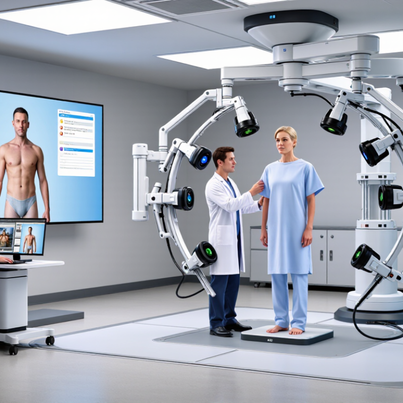
How to Choose 3D Medical Photography Systems in 2026?

What Is a 3D Medical Photography System?

A 3D medical photography system captures the patient’s body with synchronized cameras, structured light, or depth sensors. It creates a measurable three-dimensional model, not merely a flattering photograph. Clinicians can rotate the model, compare surface changes, and record standardized landmarks before treatment. The system usually includes calibrated lighting, facial or body tracking, analysis software, and secure data storage.

This matters in aesthetic surgery, dermatology, orthodontics, and reconstructive planning. The American Society of Plastic Surgeons reported nearly 1.6 million cosmetic surgical procedures in the United States in 2023. That volume increases the need for consistent documentation and clearer patient communication. A 2024 MarketsandMarkets report also identifies medical 3D imaging as a growing healthcare technology segment, driven by planning and visualization needs. Growth alone does not prove clinical value.

The image is persuasive, but it is not truth. Motion, hair, skin reflectance, lighting, and posture can distort measurements. A technician must position the patient carefully and repeat the same capture conditions. Software should show measurement accuracy, export options, access controls, and validation evidence. It should support clinical judgment, not replace it. Some systems look advanced while producing weak data. That deserves skepticism. Research quality, staff training, and transparent consent remain essential when selecting a system in 2026.

Which Clinical Applications Should the System Support?

When choosing a 3D medical photography system in 2026, clinical coverage should guide the decision. A useful system must support accurate baseline and follow-up imaging across different patient positions. It should capture facial contours, body asymmetry, scars, lesions, and wound dimensions without repeated manual measurements. Small errors matter.

In plastic and reconstructive surgery, clinicians may need preoperative planning, postoperative comparison, and realistic patient consultations. The system should document changes in volume, contour, and symmetry over time. Dermatology teams may require close imaging for lesion monitoring, while wound-care specialists need consistent records of wound size, depth estimates, and surrounding tissue changes. Burn care requires careful documentation of healing patterns. Orthopedic and rehabilitation teams may also benefit from posture, limb alignment, and movement assessments. That assumption can fail. Not every 3D system handles motion reliably.

Ask whether the platform supports standardized lighting, repeatable camera distance, and clear patient positioning instructions. These details affect clinical reliability more than impressive demonstrations. Look for measurement tools that clinicians can learn quickly. Patient review should be simple, private, and understandable. Research workflows may require exportable data, audit trails, and compatibility with existing clinical records. Privacy controls must include role-based access and secure storage. A technically advanced system can still create delays. Test it with real cases, including darker skin tones, limited mobility, and complex anatomy. If clinicians cannot use it consistently, its clinical value remains questionable. The best choice is the system that supports the applications your team performs every week, not the features that appear most advanced.

How to Compare Imaging Quality and Measurement Accuracy

How to Choose 3D Medical Photography Systems in 2026?

How to Compare Imaging Quality and Measurement Accuracy

Choosing a 3D medical photography system in 2026 requires more than impressive resolution. Imaging quality should be judged under clinical conditions, not showroom lighting. Ask for raw sample scans of skin, scars, folds, and curved body areas. Check edge definition, texture consistency, and color stability across repeated captures. Small details matter. A sharp image can still produce unreliable measurements if lighting changes between sessions. In practice, test the system on one subject three times, using a fixed position and neutral expression. The files should align without visible drift or stretched contours.

Measurement accuracy needs separate verification. Request a validation report explaining the reference method, subject range, operator training, and error calculation. Look for mean absolute error, repeatability, and confidence intervals, rather than one attractive accuracy percentage. A useful system should measure volume, distance, and surface change consistently. Test a marked mannequin or calibrated object before testing patients. Then repeat measurements after repositioning the subject. This exposes errors caused by posture, breathing, hair, or loose clothing. Results should remain clinically interpretable, not merely mathematically precise.

Workflow details also affect trust. Confirm calibration frequency, camera synchronization, software version control, and export settings. Data protection matters. Access logs and controlled storage should be documented. Involve clinicians, photographers, and measurement specialists during evaluation. Their concerns differ, and that is useful. No system is perfect. Even a validated platform may struggle with dark surfaces, reflective products, or patient movement. Record these limitations honestly, then decide whether the remaining error suits the intended clinical task.

How to Assess Workflow, Software, and Data Security

Choosing a 3D medical photography system in 2026 requires more than comparing camera resolution. The workflow must fit real clinical pressure. During a pilot, map each step from patient consent to image export. Record the time required for positioning, capture, review, and correction. A system that saves five minutes per patient can reduce daily bottlenecks.

Test the software with ordinary staff, not only technical specialists. Can users create consistent cases, compare images, and add clinical notes without confusion? Look for guided capture, automatic quality checks, role-based access, and clear audit trails. The interface should remain usable after a long clinic session. Small delays matter. Export formats also need checking, because locked data can disrupt referrals and research.

Data security deserves practical testing. Confirm encryption during transfer and storage. Ask where data is hosted, how backups are protected, and how quickly access can be removed. Review retention settings, authentication controls, and incident procedures with the provider. Request evidence of independent security assessments and documented privacy practices. Do not accept vague promises.

A test failure is useful. During evaluation, a poorly placed light caused inconsistent facial landmarks. The software flagged some images, but not all. That exposed a weakness in our review process. Build a repeatable lighting setup, train staff, and retain failed cases for review. No workflow is perfect. Continuous checks make it safer.

How to Select the Best System for Your Budget and Practice

Choosing a 3D medical photography system starts with your practice, not the display screen. Consider patient volume, available space, and the measurements you record daily. A small clinic may need a compact setup that works in one room. A busy practice may value rapid capture and automatic patient positioning. Measure twice. Hidden costs can include installation, calibration, software updates, training, and secure data storage.

Compare image consistency, depth accuracy, capture speed, and software integration. More pixels do not automatically produce better clinical data. Ask to test the system with different skin tones, body types, room sizes, and lighting conditions. Request unedited sample files, not only polished demonstrations. Check how easily staff can repeat a scan six months later. If the workflow feels complicated during a live trial, it may become slower during a busy clinic day.

Set a realistic budget based on clinical value and maintenance needs. A lower-priced system may be suitable for occasional documentation, while advanced practices may justify stronger automation and reporting tools. Confirm service response times, replacement procedures, user training, and export options before signing anything. Do not accept vague promises. Independent evaluations and references from similar practices can reveal practical weaknesses. A polished demo can hide them. My own planning mistake would be underestimating staff time; every extra minute affects appointments, staffing, and patient comfort. Reassess the choice after a trial period, because the most impressive system may not be the most dependable one.

How to Choose 3D Medical Photography Systems in 2026? - How to Select the Best System for Your Budget and Practice

Practical comparison of non-branded 3D medical photography system configurations. Prices are typical 2026 planning ranges in USD and should be confirmed through a formal quotation.
System configuration Typical purchase range Capture method Capture time Typical 3D output Recommended room area Best suited for Main limitation
Portable single-camera setup $3,000–$10,000 Controlled multi-angle photographs or smartphone-based photogrammetry 2–10 minutes, including positioning Textured surface model; usually suitable for visual comparison and basic measurements Approximately 6–10 m² Small practices, consultations, outreach clinics, and occasional documentation More sensitive to patient movement, lighting changes, camera positioning, and operator technique
Fixed dual-camera clinical kit $10,000–$25,000 Synchronized stereo or structured-light capture with calibrated lighting 30–90 seconds High-resolution textured model with repeatable landmarks and surface measurements Approximately 10–16 m² Routine before-and-after records, facial assessment, body contouring, and treatment planning Limited viewing angles may leave gaps in complex anatomy or areas with occlusion
Multi-camera clinical studio $25,000–$60,000 Synchronized multi-camera photogrammetry with fixed patient positioning and uniform illumination 1–5 seconds Full-body or regional 3D model with detailed texture, volumetric comparison, and standardized views Approximately 16–25 m² High-volume cosmetic, reconstructive, dermatology, breast, and body-contouring practices Higher installation cost, dedicated space, and stronger requirements for calibration and workflow training
Automated high-throughput studio $60,000–$150,000+ Large synchronized camera array, automated lighting, guided positioning, and centralized processing Less than 1–3 seconds Comprehensive high-resolution model with automated landmarks, serial comparison, and advanced reporting options Approximately 25–40 m² Large clinics, teaching centers, clinical research, standardized multicenter studies, and high patient volumes Highest capital cost and more demanding requirements for IT integration, privacy controls, and staff training
Specialized handheld 3D scanner $8,000–$35,000 Handheld optical scanning using structured light or laser-based surface acquisition 1–10 minutes per region Very detailed local surface geometry; texture and color capability vary by configuration Approximately 6–12 m² Small anatomical regions, custom devices, wound assessment, orthotics, and irregular surfaces Not ideal for rapid full-body capture; operator motion and patient movement can affect registration
Evaluation note: Confirm measurement accuracy on the anatomical region you document most often. Ask for repeatability testing, data-export formats, image retention policies, encryption, role-based access, audit logs, backup procedures, software subscription terms, calibration requirements, and integration with your electronic medical record system before purchase.

FAQS

Which clinical applications should a 3D medical photography system support?

It should document facial contours, body asymmetry, scars, lesions, and wounds. It should support baseline and follow-up imaging. Small errors matter. Plastic surgery, dermatology, burn care, wound care, orthopedics, and rehabilitation may need different tools.

How can clinicians evaluate imaging quality?

Request raw scans of skin, scars, folds, and curved body areas. Check edge definition, texture consistency, and color stability across repeated captures. Showroom lighting can mislead. Test under ordinary clinical conditions.

How should measurement accuracy be verified?

Ask for validation details, including reference methods, subject ranges, and error calculations. Look for mean absolute error, repeatability, and confidence intervals. Test a calibrated object before testing patients. Then repeat scans after repositioning.

Can posture and movement affect scan reliability?

Yes. Breathing, hair, loose clothing, and posture can change measurements. Some systems handle motion poorly. That assumption can fail. Use fixed positioning, consistent camera distance, and clear patient instructions.

What tests should be included before purchasing a system?

Test different skin tones, body types, room sizes, and lighting conditions. Include limited mobility and complex anatomy. Capture one subject three times. The files should align without stretched contours or visible drift.

What workflow features are important for daily clinical use?

Staff should learn measurement tools quickly. Patient review should be private and understandable. Check capture speed, positioning guidance, calibration, software updates, and export settings. A technically advanced system can still create delays.

What privacy and data controls should be checked?

Confirm role-based access, secure storage, access logs, and controlled data handling. Research teams may also need audit trails and exportable files. Privacy cannot be an afterthought.

How should a clinic compare system costs?

Include installation, calibration, training, software updates, maintenance, and secure storage. A compact setup may suit a small clinic. A busy practice may need faster capture. More pixels do not automatically mean better clinical data.

How can a clinic judge whether the system is dependable?

Request unedited samples and references from similar practices. Run a live trial with real cases. Record weaknesses honestly. My own planning mistake would be underestimating staff time. Reassess after the trial.

Conclusion

Choosing the right 3D Medical Photography Systems in 2026 requires more than comparing camera specifications. A suitable system should capture accurate, consistent three-dimensional images and support the clinical applications most relevant to a practice, such as treatment planning, progress tracking, patient education, and outcome assessment. Buyers should evaluate image resolution, color consistency, scanning speed, repeatability, and measurement accuracy under real clinical conditions.

Workflow and data management are equally important. The system should offer intuitive software, efficient image storage, flexible export options, user access controls, reliable backups, and strong protection for sensitive patient information. Practices should also consider staff training, maintenance, room requirements, integration with existing workflows, and long-term operating costs. By balancing clinical needs, imaging performance, usability, security, and total budget, healthcare providers can select a dependable solution that improves documentation and supports better-informed clinical decisions without creating unnecessary operational complexity.

Clara

Clara

Clara is a professional marketing specialist with a strong understanding of product strategy, customer needs, and digital communication. As a core member of the company’s marketing team, she combines market research, content development, and practical product knowledge to create clear, valuable,......