Knowledge & Innovation

Aesthetic Medicine & 3D Imaging Blog

Latest clinical research, technical advances, and practice growth insights from LifeViz®.

0%

Buttock Augmentation 3D Photography helps clinicians document body shape with more detail than standard two-dimensional photographs. A camera captures surface geometry, while software builds a visual model for review and measurement. The result depends on more than the device. Lighting, camera distance, patient position, and calibration can all affect the image. Small changes matter.

This guide introduces ten types of systems, including structured-light scanners, stereo-camera setups, photogrammetry platforms, and portable imaging units. Some combine several methods, so categories may overlap. A neat ranking can hide important trade-offs. For example, a fixed studio system may offer repeatable positioning, while a handheld scanner can be easier to move between rooms. Neither is automatically best for every clinic.

The comparison focuses on practical features: capture workflow, model detail, setup needs, and consistency across visits. These tools support visual documentation; they do not replace clinical assessment or guarantee a particular result. Reliable use calls for trained operators, consistent protocols, secure handling of patient images, and clear consent. Even then, measurements can vary. That limitation deserves attention. Readers should confirm technical specifications and clinical suitability with qualified professionals before choosing a system.
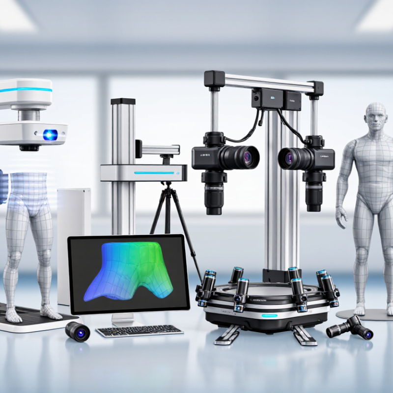
Top 10 Types of Buttock Augmentation 3D Photography Systems?

How 3D Photography Supports Buttock Augmentation Planning

Three-dimensional photography can help patients and surgeons discuss buttock augmentation with clearer visual references. A standardized set of front, side, and oblique views records the body’s surface contours. The model can be rotated to show how the buttocks relate to the waist, hips, and thighs. Small differences between sides may become easier to notice. That matters.

During consultation, a qualified surgeon can use these images to compare a patient’s goals with their existing anatomy. Digital simulations may illustrate possible changes in contour or proportion, but they are discussion tools, not promises of a result. Skin quality, health, surgical technique, and healing all influence outcomes. A rendered image cannot show internal tissue or determine whether a procedure is appropriate.

The capture itself can be imperfect. Lighting, posture, camera distance, or slight movement may alter how a contour appears, so consistent positioning is important. Images should be reviewed alongside an in-person examination and a detailed medical history. Patients should also know how their photographs are stored and who can access them. The picture helps guide a conversation; it does not replace clinical judgment.

Core Imaging Principles and Gluteal Measurements

3D buttock photography records the body’s outer surface, not its internal tissues. Systems may use structured light, paired cameras, or multiple photographs to build a three-dimensional model. Each method depends on clear visibility, stable positioning, and consistent lighting. Shadows can distort contours. So can loose clothing or a slight shift in stance.

Useful gluteal measurements include maximum width, projection from a defined side-view plane, and circumference at a repeatable level. A 3D model can also compare left-right shape and estimate surface volume within marked boundaries. These values mean little without consistent landmarks. The pelvis should remain level, feet placed evenly, and camera distance unchanged between sessions. Small details matter.

In clinical use, measurements support discussion and follow-up; they do not replace examination or determine a suitable procedure. Surface-based volume estimates are not the same as anatomical volume, and software boundaries can change the result. A careful operator should document positioning, measurement points, and limitations. I would still question any report that presents a single number as exact. Bodies move, and no capture is perfectly identical.

Top 10 Types of Buttock Augmentation 3D Photography Systems? - Core Imaging Principles and Gluteal Measurements

System Type Core Imaging Principle Potential Gluteal Measurements Practical Strengths Important Limitations
Multi-camera stereophotogrammetry Several synchronized cameras capture the subject from different viewpoints. Corresponding image features are used to reconstruct a three-dimensional surface. Surface shape, buttock width and projection, left-right surface differences, and pre- to post-procedure surface-volume change within a defined region. Captures a broad surface quickly and can reduce motion-related mismatch when cameras are synchronized. Requires calibration, consistent positioning, adequate coverage, and repeatable region boundaries. The reconstructed surface does not show internal tissue or implant position.
Structured-light 3D scanning A projector casts a known pattern onto the body; cameras record how the pattern deforms to calculate surface depth. Three-dimensional contour, regional surface area, projection from a chosen reference plane, and registered surface change. Provides dense surface detail without contact and is suitable for standardized pre- and post-treatment documentation. Movement during capture, occlusion in the gluteal cleft, reflective materials, and inconsistent stance can affect reconstruction.
Fringe-projection profilometry Sinusoidal light fringes are projected onto the surface. Phase shifts in the observed fringes are used to derive surface depth. Local curvature, contour profile, surface asymmetry, and regional shape change after surface registration. Can acquire detailed surface geometry over a defined field of view. Usually needs stable positioning and controlled lighting. Deep folds or hidden surfaces may not be captured reliably.
Laser-line triangulation scanning A laser line is projected across the body and viewed by a camera at a known angle. Triangulation converts the line position into depth. Cross-sectional contour profiles, surface distances, and shape differences along scanned paths. Can produce precise local profiles when the scanner geometry and subject position are controlled. Coverage may require multiple passes or views. Hand movement, incomplete overlap, and occluded areas can introduce errors.
Time-of-flight depth imaging The sensor estimates distance from the time delay or phase shift of emitted and returned light. Approximate three-dimensional surface shape, overall projection, and broad regional changes. Captures depth maps rapidly and can be useful for general positioning or larger-scale surface assessment. Depth resolution and edge detail vary by device and distance. Results can be affected by ambient light, surface properties, and motion.
Passive stereo vision Two or more cameras observe the subject without projected patterns. Depth is estimated by matching visible features across images. Surface contour, width, projection, and asymmetry where sufficient image texture is visible. Does not require active illumination and can use a relatively simple capture setup. Uniform skin, shadows, and occlusion can make feature matching difficult. Calibration and stable camera geometry remain essential.
Handheld RGB-D scanning A handheld device combines color images with depth data from a depth sensor; successive frames are aligned to build a surface model. Regional surface shape, contour profiles, and approximate registered surface-volume differences. Portable and flexible for capturing selected views or areas that are difficult to access with a fixed setup. Scan path, operator technique, tracking loss, and movement can affect alignment. Completeness and repeatability should be checked.
Calibrated multi-view photographic reconstruction Overlapping photographs taken from multiple angles are processed using camera geometry and image-feature matching to create a 3D surface. Overall external contour, width, projection, and surface changes when capture and scaling are standardized. Uses familiar photographic capture and can document a larger area with multiple views. Requires overlap, consistent scale, stable camera settings, and adequate visible texture. It is generally more sensitive to capture conditions than dedicated depth systems.
Single-camera structured-light scanning A camera records a projected coded or patterned light sequence; the known pattern-camera relationship is used to calculate depth. Localized buttock contour, projection, and surface profile over the scanner’s capture area. Compact configurations can capture a surface without a multi-camera array. Field of view may be limited, so multiple scans may be needed. Alignment between scans and occluded regions require particular care.
Turntable multi-view 3D surface capture Multiple views are collected as the subject or camera changes orientation; the views are registered and combined into a 3D surface. Combined external contour, regional width and projection, and surface-area or shape comparisons. Additional viewing angles can improve coverage of the sides and posterior contour. Maintaining the same posture throughout capture is challenging. Any movement between views can create alignment artifacts or distort the reconstructed surface.

Measurement note: Surface-volume change is calculated from registered 3D surfaces within a consistently defined region; it is not the same as injected fat volume or implant volume. Comparisons require standardized posture, camera or scanner calibration, scale, lighting, and anatomical landmarks. 3D surface imaging documents external shape and does not assess internal anatomy.

Ten Types of 3D Photography Systems for Buttock Assessment

Ten Types of 3D Photography Systems for Buttock Assessment

Structured-light scanners project patterns to map surface depth. Multi-camera stereophotogrammetry combines synchronized views. Handheld structured-light units capture close-range contours. Laser triangulation scanners measure surface profiles. Time-of-flight cameras estimate depth from reflected light. Fringe-projection systems read distorted light bands. RGB-D cameras pair color images with depth maps. DSLR multi-view photogrammetry builds models from overlapping photographs. Smartphone photogrammetry uses multiple images, but depends heavily on lighting and camera handling. Hybrid 2D/3D systems combine surface models with conventional photographs.

These systems can document shape, asymmetry, and changes in volume, but they do not measure tissue quality or predict surgical outcomes. Consistent posture matters. A small shift in hip rotation can alter the apparent contour. In clinical practice, repeatable lighting, camera distance, and patient position are essential, yet difficult to standardize across visits. Some systems also struggle with reflective skin, deep folds, or movement. Imperfect data needs careful interpretation.

The American Society of Plastic Surgeons’ 2023 procedural statistics recorded 28,638 buttock augmentations with fat grafting in the United States. This figure describes procedures, not 3D imaging use. It does underline why clear, comparable documentation matters during assessment and follow-up. Clinicians should select equipment based on validated accuracy, workflow, and patient privacy, rather than image sharpness alone.

Top 10 Types of 3D Imaging Systems for Buttock Assessment

A comparison of typical imaging capabilities. Values are binary: 1 = typically available; 0 = not provided by the imaging method itself.

Optical systems capture external surface shape and do not show internal anatomy. CT and MRI can depict internal structures and reconstruct the body surface; CT uses ionizing radiation, while MRI does not. These are general technical characteristics, not a ranking of clinical accuracy or suitability.

How Capture Methods and Software Differ

Buttock augmentation 3D photography systems differ in how they capture shape. Ten common configurations include single-camera photogrammetry, multi-camera photogrammetry, stereophotogrammetry, structured-light booths, handheld structured-light scanners, laser triangulation, rotating-platform rigs, fixed multi-view booths, tablet depth cameras, and hybrid photo-depth systems. Some freeze a pose in seconds; others require turning or repeated passes. Small movements matter. A heel shift can change the apparent contour.

Software creates another divide. One package may align images and build a surface mesh; another may add landmarks, measurements, or side-by-side volume comparisons. Ask whether settings remain consistent across visits, and whether users can review raw captures before smoothing. Polished images are not automatically precise. No setup is perfect. Lighting, posture, camera distance, and operator technique can all affect comparisons.

The ISAPS 2023 Global Survey estimated 34.9 million aesthetic procedures worldwide in 2023. That figure describes procedure volume, not 3D imaging accuracy or adoption. For clinical evaluation, request repeatability data from the system’s own validation tests, captured under realistic conditions. Compare the same patient position, camera distance, and lighting; document any changes. This takes discipline, and busy clinics may not get it right every time. A useful system makes those steps visible rather than hiding them behind a smooth render.

Clinical Workflow and Criteria for Choosing a System

A 3D photography system should fit the clinic’s workflow, not dictate it. Before capture, staff can explain the process, obtain informed consent, and confirm how images will be stored and accessed. Use a consistent room setup, with marked foot positions and even lighting. Lighting still matters. Patients should stand in repeatable positions, with posture and camera distance recorded where possible. Clear instructions help reduce movement between views. Small differences in stance can change the apparent contour, so images are best compared under similar conditions.

When choosing a system, assess capture time, image consistency, usable body-position options, and staff training needs. Ask whether the software can display measurements and surface changes clearly, and whether results can be exported securely into the clinical record. A smooth demonstration is not enough. Test the system with varied body shapes and realistic appointment schedules. Confirm what technical support and maintenance are available. Most importantly, treat images as one aid to discussion, not a promise of surgical results or a replacement for examination. Even careful protocols leave room for variation. That limitation deserves honest discussion with patients.

FAQS

How can 3D photography help plan buttock augmentation?

It records front, side, and angled views of body contours. A surgeon can rotate the model and discuss proportions around the waist, hips, and thighs.

Can a digital simulation show my exact surgical result?

No. It can illustrate possible changes, but it cannot promise an outcome. Healing, skin quality, health, and surgical technique all affect results.

What can 3D images reveal during an assessment?

They may make differences between the two sides easier to see. They can document surface shape and changes over time, but not tissue quality.

Do posture and lighting affect the images?

Yes. A slight hip turn, uneven lighting, or movement can change how contours appear. Consistent positioning helps.Small shifts matter.

What types of 3D photography systems are used?

Options include structured-light scanners, multi-camera systems, laser scanners, and camera-based photogrammetry. Each has different capture needs and limitations.

How should a clinic choose a system?

Consider capture time, image consistency, staff training, and secure export options. Test it with varied body shapes and realistic appointment schedules.

How should 3D images be used in consultation?

Review them alongside an in-person examination and medical history. They support discussion, but do not replace clinical judgment.

What should patients ask about image privacy?

Ask how photographs are stored and who can access them. Clear answers matter; a polished image alone is not enough.

Conclusion

This article explains how Buttock Augmentation 3D Photography can support pre-procedure planning by creating a structured visual record of gluteal shape, volume, and symmetry. It introduces core imaging principles and measurements used to describe the buttocks, such as contour, projection, proportions, and differences between the two sides. These observations can help clinicians and patients discuss goals more clearly, while 3D images remain a planning aid rather than a substitute for professional assessment.

The article also surveys ten types of 3D photography systems used for buttock assessment, comparing how their capture methods and software create, display, and analyze body images. It outlines how factors such as image consistency, measurement tools, ease of use, and integration into a clinical workflow may influence system selection. By considering both imaging capabilities and practical needs, readers can better understand how different approaches support documentation, communication, and individualized planning.

Evelyn

Evelyn

Evelyn is a professional marketing specialist dedicated to helping businesses understand products, markets, and customer needs through clear, practical, and insightful communication. With extensive knowledge of the company’s product portfolio, service processes, and industry applications, Evelyn......