Aesthetic Medicine & 3D Imaging Blog
Latest clinical research, technical advances, and practice growth insights from LifeViz®.
7 Best 3D Facial Rejuvenation Analysis Systems?
Choosing the 7 Best 3D Facial Rejuvenation Analysis systems requires more than comparing software features. It requires evidence, clinical experience, and careful interpretation. Modern platforms can map facial depth, asymmetry, skin texture, and projected volume changes within seconds. A patient may see a faint cheek shadow, a softer jaw contour, or uneven light across the nasolabial area. These details can influence treatment planning.
Demand for aesthetic care is substantial. The ISAPS Global Survey reported 34.9 million surgical and nonsurgical aesthetic procedures worldwide in 2023, including 19.1 million nonsurgical procedures. The American Society of Plastic Surgeons also recorded more than 26 million cosmetic and reconstructive procedures in its 2023 statistics. These figures support the need for consistent assessment tools, not automated promises.
Dr. Steven Dayan, a facial plastic surgeon and aesthetic medicine educator, has stated, “Beauty is not a mathematical equation.” That principle matters here. A 3D scan can measure contours, but it cannot fully measure expression, identity, cultural preferences, or patient confidence. The best systems therefore combine calibrated imaging, repeatable photography, clinician review, and transparent limitations. Research reports are useful, but they are not interchangeable. Marketing claims can also look more precise than the underlying validation.
This guide examines seven leading systems through image accuracy, workflow, visualization, data security, clinical usability, and evidence quality. Some platforms perform well in controlled lighting yet become less reliable with movement, makeup, or changing facial expressions. That weakness deserves attention. A credible 3D Facial Rejuvenation Analysis system should support informed decisions, not replace professional judgment or meaningful patient consent.
What Is 3D Facial Rejuvenation Analysis?
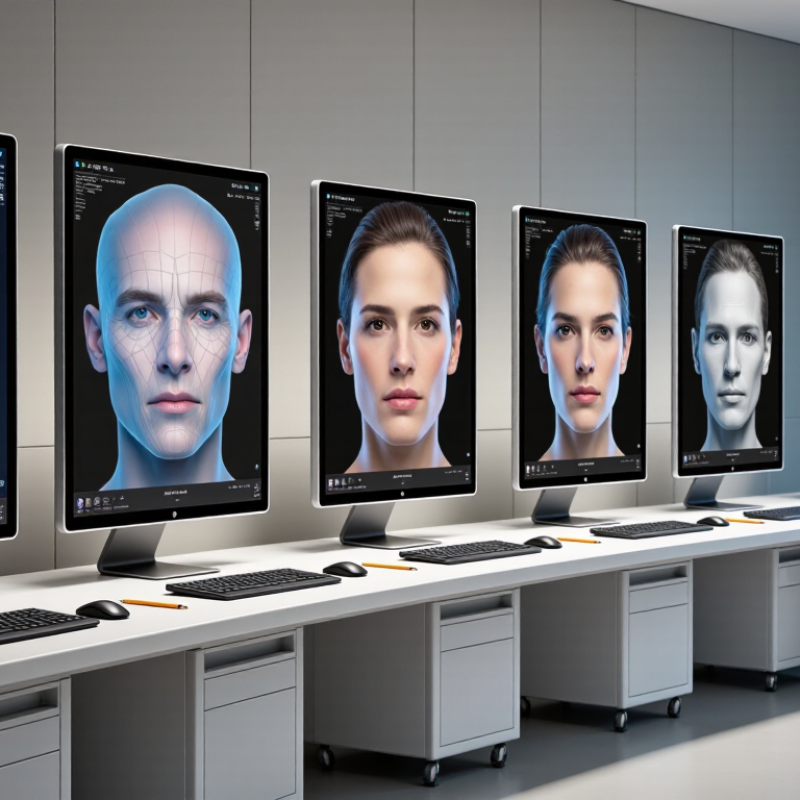
3D facial rejuvenation analysis is a clinical imaging process that maps facial shape, skin surface, and visible age-related changes. A scanner captures depth from multiple angles, creating a digital model of the face. Trained professionals may examine fine lines, volume loss, uneven contours, and facial asymmetry.
The process is more detailed than a standard photograph. It can show a shallow cheek depression, a stronger fold beside the mouth, or different cheek heights. Measurements may help guide treatment discussions and document changes over time. Consistent lighting, head position, and facial expression matter. Even a slight tilt can affect the result.
The technology has limits. It does not reveal every skin concern, and it cannot predict how a person will respond to treatment. Hydration, recent sun exposure, swelling, and camera quality may alter the images. The numbers can mislead. A qualified medical professional should interpret the findings alongside medical history, physical examination, and personal goals. In practice, comparing images under matching conditions is more useful than chasing a perfect score. I would also question dramatic improvements shown without identical lighting or neutral expressions. That detail is often overlooked.
How 3D Facial Scanning and Imaging Systems Work
7 Best 3D Facial Rejuvenation Analysis Systems?
How 3D Facial Scanning and Imaging Systems Work
A 3D facial scanning system captures depth, contours, and skin texture without touching the face. Structured light or stereo cameras project patterns across the skin. Sensors record how those patterns change. Software then builds a digital facial map within seconds. It measures areas such as cheek volume, facial symmetry, wrinkles, pores, and pigmentation. The process is usually painless. Small movements can still affect accuracy.
Clinicians compare scans from different visits using fixed landmarks around the eyes, nose, lips, and jawline. This creates a clearer view of subtle changes than ordinary photographs. The American Society of Plastic Surgeons reported millions of minimally invasive aesthetic procedures in its 2023 statistics. That demand increases the need for measurable treatment planning. ISAPS also recorded more than 19 million nonsurgical procedures worldwide in its 2023 global survey. Yet, a scan does not diagnose every skin condition. It supports professional judgment, but cannot replace it.
Tips: Use consistent lighting, camera distance, facial expression, and head position. Remove reflective makeup when possible. Ask whether the system records texture, depth, or both. Results may look impressive, but they are not perfectly objective. Facial swelling, hydration, and lighting can change the image. I would review raw images, measurement settings, and clinical photographs together. That extra step may reveal errors before treatment decisions are made.
Key Facial Aging Metrics Measured by 3D Analysis
Three-dimensional facial rejuvenation analysis turns visible aging into measurable data. The ISAPS International Survey 2023 reported more than 34 million aesthetic procedures worldwide. That growth increases the need for consistent facial assessment.
A useful system measures wrinkle depth, skin roughness, pore visibility, and pigmentation density. It also records facial volume in the cheeks, temples, lips, and jawline. Small changes matter. A 0.5-millimeter difference may influence treatment planning, although lighting can distort that result. Facial symmetry is another key metric. Software compares left and right contours, including eye height, cheek projection, and mouth position.
Perfect symmetry is not realistic.
The system should track skin elasticity and surface displacement during expression. Static images can miss movement-related folds. A 2022 review in Aesthetic Surgery Journal linked three-dimensional imaging with more objective evaluation of facial volume and contour changes. Another analysis in Clinical, Cosmetic and Investigational Dermatology emphasized standardized lighting and positioning for reliable skin comparisons. These controls are often overlooked.
Age estimation can support analysis, but it should not replace clinical judgment. Facial aging differs across genetics, sun exposure, weight change, and facial movement. The measurements look precise. They are not infallible. Professionals should compare scans over time, document device settings, and explain uncertainty before interpreting results.
Seven Leading 3D Facial Rejuvenation Analysis Systems
The demand for measurable facial assessment continues to grow. The ISAPS Global Survey 2023 recorded 8.88 million botulinum toxin procedures and 5.55 million hyaluronic acid procedures worldwide. That volume increases the need for consistent imaging before treatment. Seven leading 3D facial rejuvenation analysis systems now support clinical decisions: stereophotogrammetry, structured-light scanning, multispectral imaging, AI skin analysis, facial-landmark mapping, volumetric simulation, and longitudinal outcome tracking.
Stereophotogrammetry captures a detailed facial surface using multiple synchronized images. Structured-light scanning projects patterns onto the face and measures contour changes in millimeters. Multispectral imaging examines pigmentation, redness, and vascular patterns beneath ordinary lighting. AI skin analysis can detect texture and uneven tone, but its results depend on calibration, skin type, and image quality. Facial-landmark mapping measures symmetry around the eyes, nose, lips, and jaw. Small differences matter.
Volumetric simulation estimates how fillers or tightening procedures may alter facial balance. It should guide discussion, not promise an exact result. Longitudinal tracking compares standardized images across months, helping clinicians review swelling, tissue changes, and patient satisfaction. The American Society of Plastic Surgeons reported over 5.4 million minimally invasive cosmetic procedures in 2023, showing why repeatable measurement matters. Still, no system fully understands expression, lighting, aging, or personal expectations. A technically accurate scan can remain clinically incomplete. Human review is essential.
7 Best 3D Facial Rejuvenation Analysis Systems
Comparative capability scores across clinically relevant 3D facial analysis dimensions
The chart compares seven anonymized system profiles using a normalized 0–100 capability scale. The evaluation dimensions reflect common requirements in 3D facial rejuvenation analysis: surface mapping, wrinkle assessment, pigmentation analysis, facial symmetry, volume evaluation, and treatment-planning visualization. Higher scores indicate broader analytical functionality and stronger visualization support, not guaranteed clinical outcomes.
How to Compare Accuracy, Usability, and Clinical Value
7 Best 3D Facial Rejuvenation Analysis Systems: How to Compare Accuracy, Usability, and Clinical Value
The best system is not always the one with the sharpest image. Accuracy begins with repeatable capture, stable lighting, and consistent facial positioning. Check whether the device reports measurement error, scan-to-scan variation, and validation results from real clinical settings. NIST’s Face Recognition Technology Evaluation shows that image quality and demographic factors can influence facial analysis errors. That lesson matters here.
Look for practical workflow details. Can staff complete a scan in under two minutes? Does the software flag head movement, shadows, or incomplete landmarks? A clear audit trail is essential. It should preserve the original scan, analysis settings, and comparison date. These details support reliable consultations and reduce exaggerated promises. Still, convenience can hide weakness.
Clinical value needs stronger evidence. The ISAPS Global Survey 2023 recorded 34.9 million aesthetic procedures worldwide, including 19.1 million nonsurgical treatments. More patients now expect measurable, natural-looking changes. A useful system should connect facial measurements with treatment planning, follow-up photographs, and documented outcomes. It should not simply produce colorful heat maps. Ask whether independent clinicians tested its predictions and whether results changed decisions. I remain cautious when vendors report accuracy without defining the reference standard. A perfect score sounds impressive, but real faces rarely behave perfectly.
FAQS
Structured light or stereo cameras project patterns onto the face. Sensors record pattern changes and create a digital facial map within seconds. The scan measures depth, contours, wrinkles, pores, pigmentation, and symmetry.
No physical contact is usually required. The process is generally painless. Small movements can still reduce accuracy.
It can assess cheek volume, facial symmetry, wrinkles, pores, and pigmentation. Results depend on the system’s measurement functions. Some systems record texture, depth, or both.
Use consistent lighting, camera distance, facial expression, and head position. Remove reflective makeup when possible. Hold still.
No. A scan supports professional judgment but cannot replace clinical assessment. It may show visible patterns without explaining their medical cause.
Clinicians use fixed landmarks around the eyes, nose, lips, and jawline. They compare scans with consistent lighting and positioning. Swelling and hydration may still change the results.
Review scan-to-scan variation, measurement error, and clinical validation. Check whether the software flags shadows, head movement, or missing landmarks. Convenience can hide weakness.
An audit trail should preserve the original scan, settings, and comparison date. Reviewing raw images beside clinical photographs may reveal errors. Colorful heat maps alone are not enough.
Not always. Repeatable capture and useful treatment planning matter more than visual sharpness. A perfect score sounds impressive, but real faces rarely behave perfectly.
Conclusion
3D Facial Rejuvenation Analysis uses advanced three-dimensional scanning, imaging, and software-based modeling to create a detailed map of the face. Unlike ordinary photographs, these systems capture facial depth, contours, symmetry, volume, skin texture, and surface changes from multiple angles. By comparing images over time, practitioners can evaluate visible signs of aging, including wrinkles, folds, sagging, uneven volume, pigmentation patterns, and changes in facial proportions.
This guide reviews seven leading types of 3D facial analysis systems and explains how they differ in imaging methods, measurement detail, workflow, and reporting capabilities. It also outlines the main factors to consider when comparing accuracy, repeatability, ease of use, patient comfort, data visualization, and clinical value. The best system should provide reliable results, support objective treatment planning, make progress easy to understand, and integrate smoothly into professional consultation and follow-up processes.

Blog Tags: