Aesthetic Medicine & 3D Imaging Blog
Latest clinical research, technical advances, and practice growth insights from LifeViz®.
Why Is Skin Microstructure Analysis Important for Buyers?
Table of Contents
- Define the Target: 3 Skin Layers and 6 Fitzpatrick Phototypes
- Map Key Measures: Pores, Wrinkles, Pigmentation, and Surface Texture
- Compare Imaging: 2D Photography and 3D Surface Topography
- Check Measurement Repeatability Across All 6 Fitzpatrick Phototypes
- Separate Skin Imaging from SPF Testing: ISO 24444:2019 Sets an SPF Method
- FAQS
- Conclusion
- Related Posts
Skin Microstructure Analysis gives buyers a closer look than a surface-level photograph. It can reveal pore patterns, fine lines, texture changes, and uneven pigmentation under controlled lighting. These details help buyers compare devices, skincare services, and product claims with clearer criteria. A polished screen is not enough. Measurement quality matters.
Grand View Research valued the global skincare products market at USD 145.3 billion in 2023 and projected continued growth through 2030. This figure describes the broader skincare market, not the skin-analysis segment. Still, it shows why buyers face a crowded field of tools and promises. Dermatologist Dr. Howard Murad is often quoted as saying, “The skin is a reflection of what’s going on inside your body.” For buyers, that idea supports careful assessment, not instant conclusions.
Look for consistent lighting, repeatable capture conditions, clear explanations of what each metric means, and evidence that results are reproducible. Ask whether the system tracks the same area over time. Ask how it handles different skin tones. Small details matter. A result can look precise and still mislead if the image setup changes. Analysis also cannot replace professional diagnosis. Buyers should treat it as one input, alongside clinical evidence and user needs. That distinction is easy to overlook, and it deserves more attention.
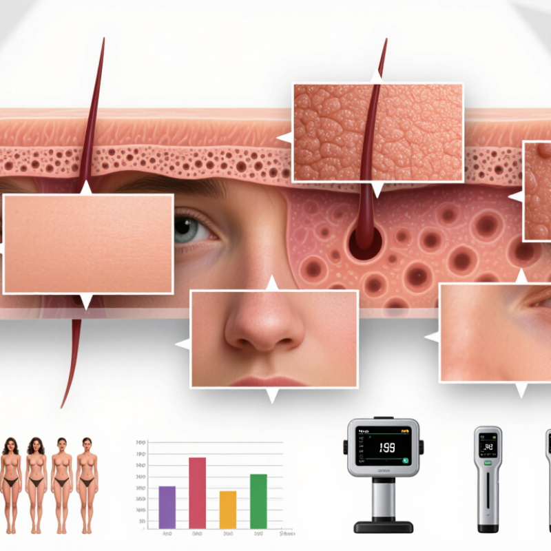
Define the Target: 3 Skin Layers and 6 Fitzpatrick Phototypes
Skin microstructure analysis helps buyers judge what a device or assessment method actually measures. The skin has three main layers: the epidermis, dermis, and subcutaneous tissue. Each has different structures and functions. A surface image may show pores or pigmentation, but it cannot automatically reveal deeper tissue conditions. That distinction matters when comparing instruments, study results, or product claims. A polished report can still leave important questions unanswered.
Fitzpatrick phototypes I through VI describe how skin tends to respond to sun exposure, especially burning and tanning. They are not a direct measure of skin color, race, or every person’s sensitivity. Two people with similar-looking skin may respond differently. For buyers, this means testing should include varied phototypes and clearly state how measurements were collected. One number rarely tells the whole story.
Tips: Ask which skin layer a tool measures, how results are validated, and whether lighting or hydration affects readings. Check that test groups include multiple phototypes. Small details matter. A protocol may look precise yet miss real-world variation. Do not treat a chart as the person.
Map Key Measures: Pores, Wrinkles, Pigmentation, and Surface Texture
Skin microstructure analysis turns visible details into comparable observations. Buyers can inspect pore appearance, wrinkles, pigmentation patterns, and surface texture instead of relying on a single overall score. Small differences matter. A close-up image may show noticeable pores around the nose, fine lines near the eyes, or uneven tone across the cheeks. Consistent lighting, camera distance, and skin preparation make these observations more reliable.
Each measure answers a different question. Pore mapping can show where openings look more noticeable, but it cannot explain why. Wrinkle measurements may distinguish fine lines from deeper folds, while pigmentation mapping highlights areas of uneven color. Texture analysis can record roughness, flaking, or visible bumps. Look for clear definitions and repeatable methods. Context matters. If the same face produces different readings under different lighting, the numbers need careful interpretation.
For buyers, useful analysis should connect measurements to practical needs, such as tracking changes or comparing areas. Ask how images are captured, whether results can be repeated, and what the system cannot measure. These tools may miss subtle changes. Human review also varies, and no image replaces clinical advice for a persistent skin concern. A precise-looking chart can still oversimplify living skin; that limitation deserves attention.
Compare Imaging: 2D Photography and 3D Surface Topography
For buyers comparing skin-analysis systems, 2D photography and 3D surface topography answer different questions. A 2D image records color and visible features, such as redness or pigmentation, but lighting can make pores and fine lines appear deeper or softer. 3D imaging adds height information, mapping raised and indented areas across the skin. That extra layer can support more consistent texture comparisons, though it does not make every assessment automatically accurate.
The EEMCO guidance on skin topography emphasizes standardized measurement conditions, while ISO 25178 defines areal surface parameters used in 3D measurement. These include Sa, the mean height deviation, and Sz, the height range between peaks and valleys. Buyers should check whether a system reports such metrics, saves repeatable images, and controls lighting and head position. Small setup changes matter. A glossy cheek under a different light can confuse a comparison.
Tips: Ask for a live demonstration using the same subject, lighting, and camera position. Compare the 2D image with the 3D height map, then ask how repeatability is checked. A polished display is not proof of reliable measurement; that part deserves scrutiny.
| Comparison Dimension | 2D Photography | 3D Surface Topography | Why It Matters to Buyers |
|---|---|---|---|
| Data captured | A flat image records pixel values, such as color and brightness, from the skin surface. | A surface map records height variations across the measured area, typically alongside a rendered image. | Choose according to whether the goal is visual documentation, surface-shape measurement, or both. |
| Texture and topography | Texture can be assessed visually or with image-analysis methods, but the image alone does not directly encode surface height. | Provides topographic information that can help quantify features such as roughness, wrinkle depth, and surface relief. | 3D data can support more direct geometric comparisons when surface change is a key outcome. |
| Color and visible appearance | Well-controlled photography is useful for documenting visible features such as redness, pigmentation, and overall appearance. | Some systems also capture color, but color capability and fidelity vary by device and acquisition setup. | For color-focused assessment, check lighting control, color calibration, and image consistency. |
| Repeatability requirements | Results depend on consistent lighting, camera settings, distance, angle, focus, and skin preparation. | Results depend on consistent positioning and acquisition settings, as well as the instrument’s calibration and measurement method. | Ask for repeatability data under the intended workflow, not just best-case specifications. |
| Sensitivity to movement | Movement during exposure can cause blur or change the apparent position of features. | Movement during capture can distort or reduce the quality of the reconstructed surface, depending on the system. | Capture speed, subject comfort, and positioning aids matter in clinical, research, and retail settings. |
| Typical outputs | Photographs, color measurements, and image-based scores or feature measurements, depending on the software. | Surface maps, height profiles, and derived topographic measurements, depending on the device and analysis software. | Confirm that output formats and metrics are suitable for reporting, longitudinal tracking, and existing workflows. |
| Equipment and workflow | Camera-based setups can be relatively straightforward, but controlled illumination and standardized positioning are important. | Requires a 3D-capable measurement system and a workflow suited to surface capture and analysis. | Compare total cost, training, maintenance, capture time, and software needs—not only the instrument price. |
| Common limitations | Shadows, reflections, lighting variation, and camera geometry can affect image-based interpretation; a flat image does not directly measure depth. | Performance can depend on capture conditions, reconstruction method, surface properties, and the region being measured. | Request validation information for the specific skin features, population, and use case you plan to assess. |
| Best suited to | Visual records, appearance assessment, and applications where color or visible change is the main focus. | Applications where surface shape, relief, or quantified topographic change is important. | The right method depends on the study question; some workflows benefit from complementary 2D and 3D data. |
| Buyer due diligence | Review lighting controls, image resolution, color calibration options, and repeatability across operators and visits. | Review measurement range, spatial sampling, height-measurement performance, calibration procedures, and analysis options. | Request specifications, validation evidence, sample reports, and a demonstration using representative conditions. |
| Measurement capabilities vary by device, software, protocol, and application. Confirm specifications and validation for the exact system and intended use before purchase. | |||
Check Measurement Repeatability Across All 6 Fitzpatrick Phototypes
Skin microstructure analysis can help buyers judge whether a device produces stable, useful measurements—not just attractive images. Test repeatability across all six Fitzpatrick phototypes, which describe how skin tends to respond to ultraviolet exposure. They are not a complete measure of skin tone or a substitute for direct assessment. Pigmentation, lighting, and camera settings can affect some image-based readings, so results from one group should not be assumed to represent everyone.
Use a consistent protocol. Keep room lighting, camera distance, positioning, and skin preparation the same. Repeat scans on the same area, then compare the readings for each phototype separately. Check whether small changes in placement produce large changes in the result. Look for sample sizes, device settings, and reported variation; a single demonstration is weak evidence. Real-world testing is rarely perfect.
Tips: Ask for repeat measurements from every phototype, not only pooled averages. Compare within-person variation as well as group averages. Record conditions. If the method has not been tested on a group, treat its performance there as uncertain rather than proven.
Separate Skin Imaging from SPF Testing: ISO 24444:2019 Sets an SPF Method
Skin microstructure images can show fine lines, pores, roughness, and visible changes in the skin surface. They help buyers compare appearance under consistent lighting and magnification. But an image cannot establish how well a sunscreen prevents sunburn. Different question. Different evidence.
ISO 24444:2019 describes an in vivo method for determining sun protection factor, or SPF. Trained personnel apply a measured amount of product to defined skin areas, then expose those areas to controlled ultraviolet radiation. They compare the amount of radiation needed to produce visible redness on protected and unprotected skin. The resulting ratio provides the SPF value under the test conditions. Small details matter: application amount, exposure settings, and assessment timing can affect results.
For buyers, this means asking for an SPF report based on the relevant method, rather than treating attractive skin images as proof of protection. Check that the tested formula matches the product being considered, and review the report’s stated conditions and results. ISO testing adds useful structure, but it cannot reproduce every climate or real-world application habit. Even careful testing has limits. A person may apply less product, miss a spot, or swim before reapplying; that gap deserves attention.
Why Is Skin Microstructure Analysis Important for Buyers?
Separate Skin Imaging from SPF Testing: ISO 24444:2019 Sets an SPF Method
This capability map distinguishes the methods, not their performance. Skin imaging can assess visible surface features such as wrinkles, pores, and texture, depending on the imaging method. ISO 24444:2019 specifies an in vivo method for determining SPF based on erythemal response. Skin imaging alone does not establish an SPF value.
FAQS
The epidermis, dermis, and subcutaneous tissue have different structures and functions. A surface image cannot automatically show deeper conditions.
They describe how skin tends to burn and tan after sun exposure. They do not fully measure skin tone or individual sensitivity.
People with similar-looking skin may respond differently. Testing across all six phototypes helps reveal where results may vary.
Scan the same area more than once. Keep lighting, camera distance, positioning, and skin preparation consistent. Record each condition.
Pigmentation, lighting, camera settings, and hydration may affect results. Small changes matter.
Not automatically. Ask which layer the tool measures and how its results were validated.
Look for sample sizes, device settings, and reported variation for each phototype. One demonstration is weak evidence.
Group averages can hide differences between people. Compare repeated readings within each person, too. Real-world testing is rarely perfect. A chart may still miss variation.
Conclusion
Skin Microstructure Analysis helps buyers understand what a skin-imaging system measures and how useful its results may be. A clear assessment should define its target across the skin’s three layers and account for all six Fitzpatrick phototypes. It should also identify the key features being evaluated, including pores, wrinkles, pigmentation, and surface texture, rather than relying on a vague overall skin score.
Imaging methods can provide different kinds of information: 2D photography records visible appearance, while 3D surface topography captures contours and relief. Buyers should check that measurements are repeatable across different skin tones and phototypes, since consistency is essential for meaningful comparisons. Skin imaging should also be distinguished from SPF testing. ISO 24444:2019 specifies an SPF test method; it does not, by itself, describe a method for analyzing skin microstructure.

Blog Tags: